1. Introduction
Endometriosis is the presence of endometrium-like tissue outside the uterus, accompanied by a chronic inflammatory reaction. It affects a substantial minority of women of reproductive age and is associated with dysmenorrhoea, chronic pelvic pain, dyspareunia and subfertility (Zondervan et al., 2020). The theory of retrograde menstruation set out by Sampson (1927) still frames most accounts of its origin, although it does not explain why only some women develop disease. Three phenotypes are conventionally distinguished: superficial peritoneal lesions, ovarian endometrioma, and deep endometriosis that infiltrates beneath the peritoneum into structures such as the uterosacral ligaments, rectovaginal septum, bowel and bladder (Zondervan et al., 2018).
The interval between the onset of symptoms and diagnosis is long. A survey conducted in the 1990s found delays of many years in both the United States and the United Kingdom (Hadfield et al., 1996), and a later study across ten countries reported a similar picture together with a measurable loss of work productivity (Nnoaham et al., 2011). Several causes contribute, including the normalisation of menstrual pain by patients and clinicians. One cause is structural. If a diagnosis can only be made at operation, it will be postponed until symptoms are judged severe enough to justify one.
This review asks what the published literature says about diagnosing endometriosis without laparoscopy. It covers clinical assessment, imaging and biomarkers, and it gives particular attention to the quality of the evidence, because the question of whether a test works cannot be separated from the question of what it was compared against.
2. Scope and Approach
This is a narrative review. It draws on the Cochrane diagnostic test accuracy reviews of imaging and biomarkers, current guidelines from European and British bodies, consensus statements on imaging technique, and primary studies that are widely cited in those sources. No formal search with screening counts was undertaken and no pooled estimates are calculated here. Where numerical accuracy figures exist they vary considerably between studies, and the review describes the direction and consistency of findings in words instead of quoting single values that could mislead.
3. Laparoscopy as an Imperfect Reference
Laparoscopy became the reference standard because it allows direct inspection of the peritoneal surfaces and biopsy of what is seen. Its accuracy is nonetheless limited. A systematic review of studies comparing visual diagnosis with histology found that a negative laparoscopy is reliable for excluding disease, whereas a positive visual impression without biopsy is less dependable (Wykes et al., 2004). Lesions vary widely in appearance, recognition depends on the surgeon's experience, and deep disease lying beneath adhesions or in the retroperitoneum may be missed on inspection alone.
Surgical staging also relates poorly to what patients experience. The revised classification of the American Society for Reproductive Medicine assigns points for lesion size and adhesions, yet a large multivariate analysis found only a weak association between stage and the severity of pain (Vercellini et al., 2007). Newer descriptive systems give more room to deep disease and can be applied to imaging as well as to surgical findings, but they too describe anatomy and not experience. These points matter for non-invasive testing in two ways. An index test can never appear better than the reference it is judged against, and a test that predicts the surgical stage is not thereby shown to predict symptoms or response to treatment.
4. Clinical Assessment
A structured history remains the first diagnostic step. Cyclical pain, dysmenorrhoea that disrupts daily activity, deep dyspareunia, cyclical bowel or bladder symptoms and subfertility each raise the probability of disease, and examination may reveal nodularity, tenderness or a fixed uterus. None of these findings is specific. Irritable bowel syndrome, bladder pain syndrome, adenomyosis and pelvic floor dysfunction produce overlapping complaints and often coexist with endometriosis, which is one reason why symptom questionnaires proposed as screening tools have not performed well enough to stand alone. Agarwal et al. (2019) argued that a clinical diagnosis based on symptoms, examination and imaging should be sufficient to begin treatment, and that insistence on surgical confirmation harms patients by delaying care. Their call to action has been widely taken up in later commentary.
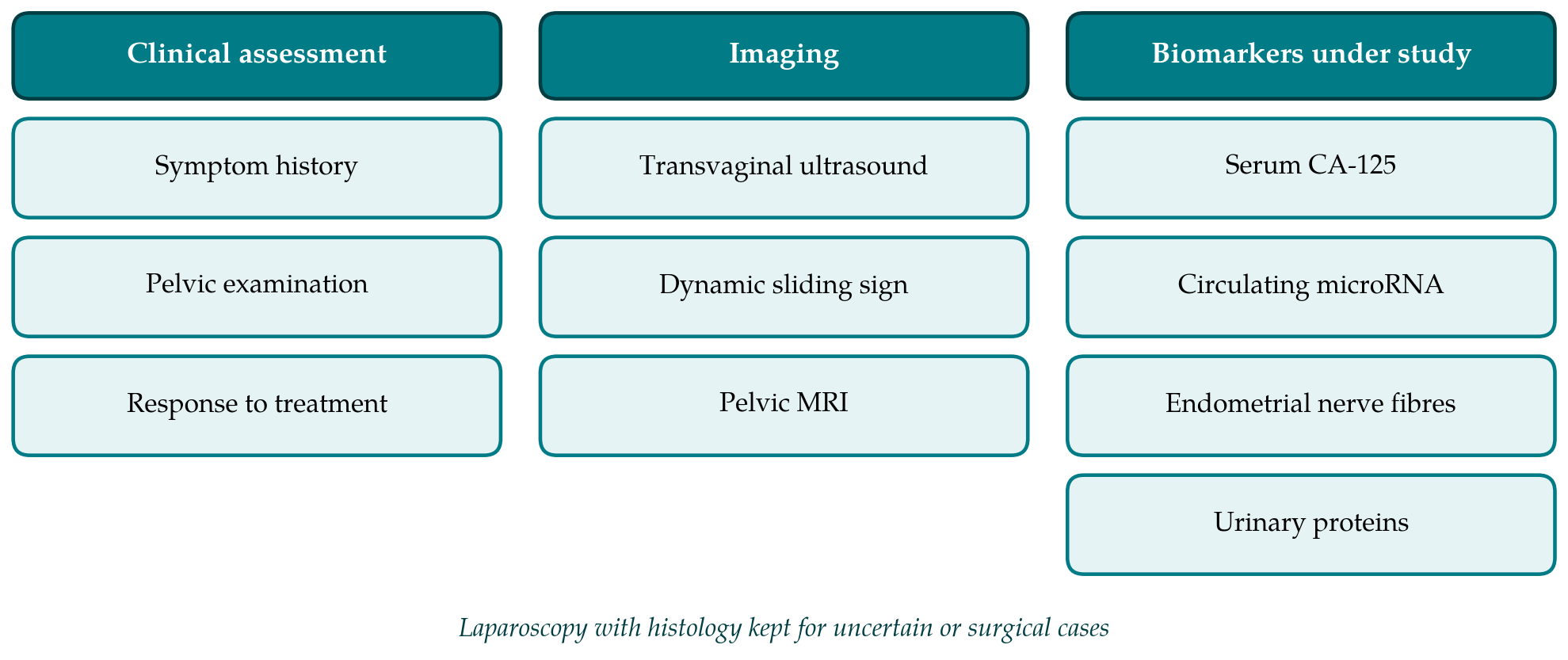
Guidelines have moved in the same direction. The 2022 guideline of the European Society of Human Reproduction and Embryology no longer treats laparoscopy as the diagnostic gold standard and recommends it only when imaging is negative or empirical treatment has failed or is inappropriate (Becker et al., 2022). The guideline of the National Institute for Health and Care Excellence likewise advises that endometriosis should not be excluded on the basis of normal examination or imaging, and supports starting treatment on clinical suspicion (NICE, 2017). Figure 1 groups the non-surgical sources of diagnostic information that these documents discuss.
5. Imaging
Imaging is the non-invasive approach with the strongest evidence. Its performance differs sharply by phenotype, and any statement about accuracy needs to specify which form of disease is meant.
5.1. Transvaginal ultrasound
Transvaginal ultrasound is the first-line imaging test. The typical endometrioma is a unilocular cyst with homogeneous low-level echoes, often described as ground glass, in a premenopausal woman, and its features have been characterised in a large multicentre series (Van Holsbeke et al., 2010). The Cochrane review of imaging concluded that transvaginal ultrasound approaches the accuracy needed to replace surgery for the diagnosis of endometrioma and performs well for deep disease of the rectosigmoid, with less certainty at other sites (Nisenblat et al., 2016a). An earlier meta-analysis reached a similar view for bowel endometriosis specifically (Hudelist et al., 2011).
Results depend on technique. The International Deep Endometriosis Analysis group published a consensus on how the examination should be performed and reported, in four steps: routine assessment of the uterus and adnexa, evaluation of soft markers such as site-specific tenderness and ovarian mobility, assessment of the pouch of Douglas using the sliding sign, and a search for deep nodules in the anterior and posterior compartments (Guerriero et al., 2016). The sliding sign, a real-time observation of whether the rectum glides freely over the posterior cervix and uterus, is used to predict obliteration of the pouch of Douglas. An examination of this kind takes longer than a routine scan and requires training that is not uniformly available. Published accuracy figures come largely from expert centres, and it should not be assumed that they transfer to general gynaecological or radiology departments where deep disease is seen less often. The consensus statement is partly an attempt to close that gap by standardising what is looked for and how it is described.
5.2. Magnetic resonance imaging
Magnetic resonance imaging is generally used as a second-line test, particularly when deep disease is suspected and surgery is being planned. It is less operator dependent at the point of acquisition, offers a wide field of view and shows sites that ultrasound reaches poorly, such as the upper sigmoid and the ureters. The European Society of Urogenital Radiology has issued recommendations on patient preparation, sequences and reporting (Bazot et al., 2017). Comparative reviews find that expert ultrasound and magnetic resonance imaging perform similarly for most deep sites and that the choice often depends on local expertise (Bazot and Daraï, 2017). Table 1 summarises how the main non-invasive options perform for each phenotype according to these sources.
| Option | Phenotype best served | Conclusion of published syntheses | Main limitation |
|---|---|---|---|
| Transvaginal ultrasound | Endometrioma; rectosigmoid deep disease | Accurate enough to guide treatment Nisenblat et al., 2016a | Operator training; misses superficial disease |
| Pelvic MRI | Deep disease, surgical mapping | Comparable to expert ultrasound Bazot and Daraï, 2017 | Cost and access; misses superficial disease |
| Serum CA-125 | Advanced disease | Too insensitive for diagnosis Mol et al., 1998 | Raised in many other conditions |
| Other blood markers | None established | No marker meets diagnostic criteria Nisenblat et al., 2016b | Small, unreplicated studies |
| Endometrial markers | None established | Promising but unconfirmed Gupta et al., 2016 | Requires an endometrial biopsy |
| Urinary markers | None established | Insufficient evidence Liu et al., 2015 | Few studies, varied methods |
| MicroRNA panels | Under investigation | Awaiting independent validation | Signatures differ between studies |
5.3. The gap: superficial peritoneal disease
Neither modality reliably detects superficial peritoneal lesions, which are small, flat and often the only form of disease present, especially in younger women. The Cochrane review found no imaging test with adequate accuracy for this phenotype (Nisenblat et al., 2016a). The practical consequence is asymmetry. A positive scan showing an endometrioma or a deep nodule can be acted on. A negative scan cannot be taken as reassurance, and guidelines say so explicitly (Becker et al., 2022). It is this gap that has sustained the search for a biomarker.
6. Biomarkers
A useful biomarker would be measurable in an accessible sample, would detect disease across phenotypes including the superficial form, and would distinguish endometriosis from other causes of pelvic pain. The Cochrane reviewers proposed explicit accuracy thresholds for a test intended to replace surgery and for a triage test intended to select who needs it, and judged each candidate against them.
6.1. Blood markers
CA-125 is the most studied marker. A meta-analysis more than two decades ago showed that its serum concentration is raised in some women with endometriosis, mainly those with advanced disease, but that its sensitivity is too low for it to serve as a diagnostic test (Mol et al., 1998). The marker is also raised in ovarian malignancy, pelvic infection, fibroids and during menstruation. A systematic review of peripheral biomarkers examined cytokines, angiogenic factors, hormones, autoantibodies and others, and found none that was clinically useful, with most reported in single small studies (May et al., 2010). The Cochrane review of blood biomarkers, which assessed a very large number of candidates, came to the same conclusion and noted the low methodological quality of the underlying studies (Nisenblat et al., 2016b).
6.2. Endometrial and urinary markers
The eutopic endometrium of women with endometriosis differs from that of unaffected women in gene expression, hormone response and innervation. Small unmyelinated nerve fibres were reported in the functional layer of the endometrium in affected women and proposed as a diagnostic sign obtainable by office biopsy (Tokushige et al., 2006). The Cochrane review of endometrial biomarkers regarded nerve fibre markers as among the more promising candidates but concluded that the evidence was insufficient to recommend any endometrial test in practice (Gupta et al., 2016). An endometrial biopsy is also less invasive than laparoscopy without being non-invasive in any ordinary sense. The review of urinary biomarkers identified too few studies of adequate quality to draw conclusions (Liu et al., 2015).
6.3. MicroRNA and other molecular signatures
Interest has shifted to panels of circulating or salivary microRNAs analysed with machine learning methods. A salivary signature reported from a French prospective cohort attracted wide attention for its apparent accuracy (Bendifallah et al., 2022). Such results should be read with the history of the field in mind. Signatures derived from high-dimensional data in a single cohort are prone to overfitting, the microRNAs identified by different groups overlap little, and pre-analytical handling affects measured levels. Recent overviews treat these tests as investigational until they are reproduced in independent populations that resemble those in which the test would be used (Taylor et al., 2021; Becker et al., 2022).
7. Methodological Problems and Research Needs
Several weaknesses recur across the diagnostic literature. The first is spectrum bias. Most studies recruit women already scheduled for surgery in specialist centres, among whom disease is common and often advanced. A test that separates severe disease from healthy pelvises in that setting may perform much worse among women with early symptoms in primary care, which is where a non-invasive test is most needed. The second is verification. Women with negative index tests are less likely to undergo laparoscopy, and when they do, the reference is itself imperfect for the reasons discussed above.
The third is reporting. Many biomarker studies select their thresholds from the same data used to estimate accuracy and omit the information that the STARD statement asks for, such as how participants were sampled and whether readers of the index test were blind to the reference result (Bossuyt et al., 2015). Future work would be strengthened by prespecified thresholds, validation in separate cohorts, recruitment at the point of first presentation, and outcomes that matter to patients, including time to treatment and symptom relief, alongside agreement with surgery. Whether a test should be judged by its prediction of visible lesions or by its prediction of who benefits from treatment is a conceptual question that the field has not settled (Taylor et al., 2021).
8. Conclusion
Endometriosis can often be diagnosed without an operation. Ovarian endometrioma and deep disease are identified by transvaginal ultrasound or magnetic resonance imaging with sufficient confidence to plan treatment, provided the examiner is trained, and current guidelines accept a clinical diagnosis as a basis for starting therapy. The same cannot yet be said of superficial peritoneal disease, for which imaging is insensitive and no biomarker has been validated. Laparoscopy therefore keeps a place, though a narrower one than it held, and the priority for research is rigorous independent evaluation of candidate tests in the populations that stand to gain from them.
Conflict of interest
To be declared by the author(s) before publication.
Funding
To be declared by the author(s) before publication.