1. Introduction
In 1972 Hoyer and colleagues described children whose nephrotic syndrome returned soon after kidney transplantation, and drew the inference that still frames the subject: something outside the kidney, and present in the recipient, must be injuring the glomerulus (Hoyer et al., 1972). Half a century later recurrent focal segmental glomerulosclerosis (FSGS) remains one of the most difficult problems in transplant nephrology. It affects a substantial minority of recipients whose native disease was primary FSGS, it can declare itself within the first days, and it shortens graft survival (Ponticelli, 2010; Cravedi et al., 2013).
Living donor transplantation sharpens every aspect of the problem. The operation is elective, the donor is a healthy volunteer, and donor and recipient are often relatives. The transplant team must decide whether the risk of recurrence is acceptable, how to explain it to two people instead of one, and whether anything can be done beforehand to lower it. This review examines the published evidence on very early recurrence with those decisions in view.
2. Approach
The article is a narrative review of the published literature on recurrence of FSGS after kidney transplantation, with attention to onset in the first days and to living donation. It draws on the original reports that established the circulating factor hypothesis, on cohort studies of risk and outcome, on reports of therapeutic interventions, and on international guidance. No systematic search was undertaken and no new data are presented. Because the therapeutic literature consists almost entirely of uncontrolled series, the review describes what has been tried and how the results have been interpreted, and does not rank treatments or recommend regimens.
3. The Clinical Picture of Very Early Recurrence
The typical presentation is proteinuria in the nephrotic range appearing soon after the graft begins to function. In the most rapid cases protein is detectable in the first urine passed. Interpretation is complicated when the native kidneys are still in place and still leaking protein, which is one reason a pre-transplant measurement of urinary protein is valuable as a baseline. Graft function may be normal at first. If proteinuria persists, oedema, hypoalbuminaemia and a progressive decline in function follow.
A biopsy taken in the first days usually looks normal by light microscopy, since segmental sclerosis takes weeks to form. The diagnostic finding is on electron microscopy: widespread effacement of podocyte foot processes, the same lesion seen in minimal change disease. The name of the disease therefore describes a late consequence. What recurs in the first week is a diffuse podocyte injury, and the scars that give FSGS its name appear only if that injury continues (D'Agati et al., 2011). The morphological variant seen in the native kidney does not reliably repeat itself in the graft.
4. Evidence for a Circulating Factor
Several observations point to a factor carried in the blood. Proteinuria can begin too quickly for any process intrinsic to the graft. Plasma from affected patients increases the albumin permeability of isolated glomeruli in a laboratory bioassay, and the effect was strongest in patients whose disease recurred (Savin et al., 1996). Removing plasma proteins by adsorption on a protein A column lowers proteinuria for as long as the treatment continues (Dantal et al., 1994), and plasma exchange has a similar effect (Artero et al., 1994). Most striking is the report by Gallon et al. (2012) of a kidney that developed recurrence within days, was removed and placed in a second recipient with a different kidney disease, and there recovered. A graft injured by the first recipient's circulation healed once it left it.
Identifying the factor has proved much harder than demonstrating its existence. Wei et al. (2011) proposed soluble urokinase plasminogen activator receptor, reporting higher serum levels in FSGS and a podocyte-activating effect in experimental models. Later studies found that the marker rises with falling kidney function and with inflammation of any cause and does not separate FSGS from other proteinuric diseases well, and most authors no longer regard it as the cause (Kopp et al., 2020). A panel of autoantibodies measured before transplantation, with anti-CD40 the most informative, was reported to predict recurrence (Delville et al., 2014) but has not entered routine use.
The most recent candidate is an autoantibody against nephrin, a structural protein of the slit diaphragm between podocyte foot processes. Watts et al. (2022) found such antibodies in a proportion of patients with minimal change disease, and a larger study confirmed their presence in minimal change disease and in some primary FSGS, with levels that tracked disease activity (Hengel et al., 2024). A Japanese multi-institutional study then detected anti-nephrin antibodies in patients with post-transplant recurrence (Shirai et al., 2024). These findings offer a plausible mechanism for at least a subset of early recurrences. Whether a pre-transplant antibody test can predict recurrence, and in what fraction of patients, has yet to be established in prospective cohorts.
5. Who Is at Risk
The single most useful step in estimating risk is to establish what kind of FSGS the candidate has, because the lesion is a pattern of injury with several distinct causes (Rosenberg and Kopp, 2017). Figure 1 sets out the main categories and what each implies. De Vriese et al. (2018) describe a practical approach: primary disease typically presents with full nephrotic syndrome and diffuse foot process effacement, while secondary or maladaptive forms, arising from reduced nephron mass, obesity, viral infection or drugs, usually show proteinuria without hypoalbuminaemia and only segmental effacement. Recurrence is essentially a feature of the primary form.
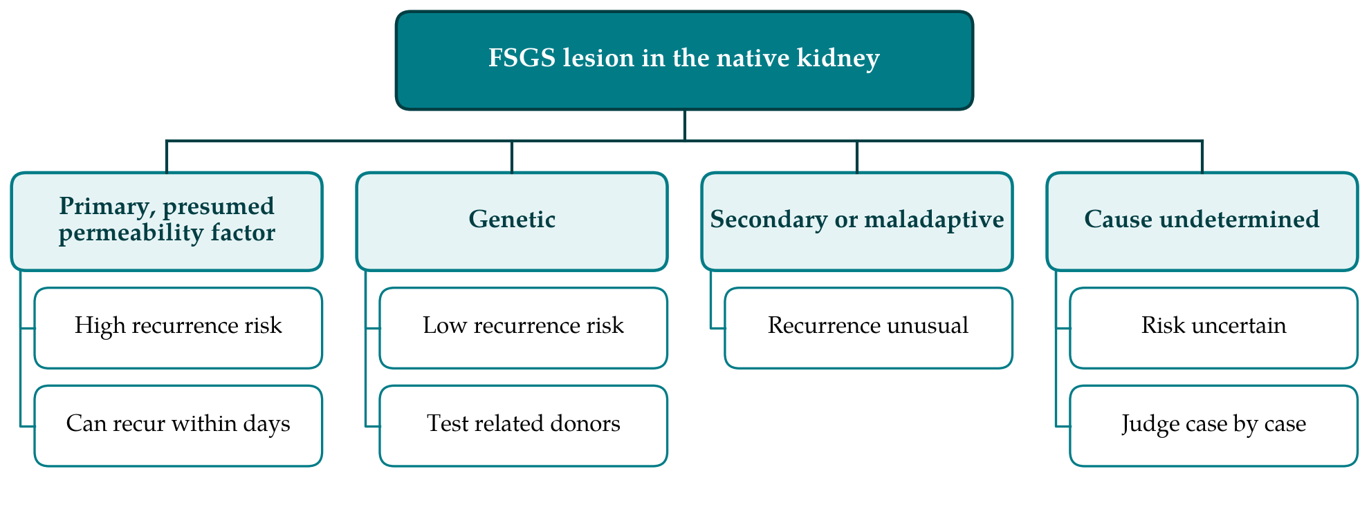
Genetic forms behave differently. Where disease results from pathogenic variants in genes encoding podocyte proteins, such as NPHS2, the defect lies in the native kidney and a graft with normal podocytes is not expected to be affected; recurrence was infrequent in a large genetically characterised series (Weber et al., 2004). Genetic testing of candidates with early-onset or steroid-resistant disease, or a family history, is therefore informative for transplant planning. Kidney disease associated with high-risk APOL1 genotypes (Genovese et al., 2010) is also intrinsic to the kidney, and here the genotype of the donor matters more than that of the recipient.
Within primary FSGS, the features most consistently associated with recurrence in older series are onset in childhood, rapid progression from diagnosis to kidney failure, and above all recurrence in a previous graft (Ponticelli, 2010). An international cohort of adult recipients found recurrence in roughly a third and reported a partly different set of associations (Uffing et al., 2020), which illustrates how unstable clinical predictors are across populations. None is accurate enough to rule recurrence in or out for an individual.
6. Questions Specific to Living Donation
Whether a living donor kidney is more likely to suffer recurrence than a deceased donor kidney has been debated for years. Published series do not show a consistent difference, and the usual advantages of living donation, including shorter time on dialysis and better graft quality, still apply. International guidance on candidate evaluation does not treat primary FSGS as a bar to transplantation, but it asks that the risk of recurrence be considered and discussed with the candidate, particularly where an earlier graft was lost to recurrence (Chadban et al., 2020). Loss of a previous graft in that way is generally regarded as a strong reason to hesitate before accepting another living donor.
Related donors raise the question of shared inheritance. If the recipient's disease is genetic, a parent or sibling may carry the same variant, and for recessive conditions a heterozygous carrier may be acceptable as a donor while an affected relative is not. Genetic evaluation of the recipient before donor work-up begins avoids discovering this late. Living donation has one clear practical advantage: the date is known. That allows baseline proteinuria to be documented, native kidney protein loss to be taken into account, monitoring to be arranged from the first post-operative day, and any pre-emptive treatment the centre uses to be scheduled.
7. Prevention and Treatment
No randomised controlled trial has tested any treatment for recurrent FSGS. Practice rests on case series, registry analyses and mechanistic reasoning, and the approaches that have been reported are summarised in Table 1. The table describes the state of evidence and is not a treatment recommendation.
| Intervention | Rationale | State of the evidence |
|---|---|---|
| Plasma exchange | Removes the presumed circulating factor | Uncontrolled series; remission in a proportion (Artero et al., 1994) |
| Immunoadsorption | Removes immunoglobulin-associated factor | Proteinuria falls during treatment (Dantal et al., 1994) |
| Rituximab | B cell depletion; possible direct podocyte effect | Series, often combined with plasma exchange (Fornoni et al., 2011) |
| Intensified calcineurin inhibitor | Immunosuppression and podocyte stabilisation | Pilot series with plasma exchange (Canaud et al., 2009) |
| Abatacept | Blocks B7-1 on injured podocytes | Initial report not confirmed (Delville et al., 2016) |
| Pre-transplant prophylaxis | Lowers factor before the graft is exposed | Conflicting observational reports; benefit unproven |
| Renin-angiotensin blockade | Lowers proteinuria non-specifically | Supportive care extrapolated from native disease |
Plasma exchange is the most widely used intervention, started promptly once recurrence is recognised. Observational series report complete or partial remission in a considerable share of patients, and cohort data suggest that grafts in which proteinuria remits last longer than those in which it does not (Hickson et al., 2009; Uffing et al., 2020). Without controls it is impossible to say how many of these remissions would have occurred anyway, or to compare schedules. Some patients become dependent on continued sessions.
Rituximab entered use after reports of remission in recurrent disease, and Fornoni et al. (2011) proposed that it might act on podocytes directly as well as on B cells. The discovery of anti-nephrin antibodies gives B cell depletion a clearer rationale in the subset of patients who carry them. Combined use with plasma exchange has been described in several series, as has an intensive regimen built around a calcineurin inhibitor (Canaud et al., 2009). The history of abatacept is a caution: an initial report of remission in patients whose podocytes stained for B7-1 (Yu et al., 2013) was not reproduced when others tested the same approach (Delville et al., 2016).
Prophylactic plasma exchange or rituximab before a planned living donor transplant is attractive in principle and feasible in practice, yet the published comparisons are small, non-randomised and inconsistent. Centres differ in whether they offer it and to whom.
8. Gaps and Future Work
Three needs stand out. The first is a validated predictive test. If anti-nephrin antibody measured before transplantation proves to identify candidates at high risk, it would change counselling for living donors and would allow preventive treatment to be studied in those most likely to benefit. The second is controlled evidence on treatment. The condition is uncommon and outcomes vary, so trials will need multicentre collaboration and agreed definitions of recurrence and remission. The third is better description of the earliest days: systematic protocols for timed proteinuria measurement and early electron microscopy would show how often recurrence begins before it is clinically obvious.
9. Conclusion
Recurrence of FSGS within days of transplantation is best understood as an immediate injury to the podocytes of a healthy graft by something in the recipient's circulation. The evidence for that mechanism is strong, and anti-nephrin autoantibodies are the most promising explanation yet for part of it. Risk depends heavily on whether the original disease was primary, genetic or secondary, which makes careful classification before transplantation the most valuable step currently available. Treatment remains empirical. For living donor transplantation the practical consequences are thorough evaluation of the recipient's diagnosis, honest counselling of both parties, and close monitoring from the first day.
Conflict of interest
To be declared by the author(s) before publication.
Funding
To be declared by the author(s) before publication.