1. Introduction
Thyroid nodules are common and most are benign. The task of fine-needle aspiration is to pick out the minority that need surgery, and it has done this well enough to become the procedure of choice in clinical guidelines (Haugen et al., 2016). An early appraisal by Gharib and Goellner (1993) established its accuracy and its effect in reducing unnecessary thyroidectomy, and reporting has since been standardized by the Bethesda System for Reporting Thyroid Cytopathology, which sorts every aspirate into one of six categories with an implied risk of malignancy and a recommended action (Cibas and Ali, 2009; Ali et al., 2023).
None of this works unless the slide is readable. For most of the history of the technique the slide was a direct smear, made by whoever held the needle. Liquid-based cytology offers another route: the sample goes into a vial of fixative and the laboratory prepares the slide. The method removes a notoriously variable manual step, and it adds cost and changes the picture under the microscope. Opinions on whether the exchange is worthwhile for the thyroid have differed sharply (Ljung, 2008; Fadda and Rossi, 2011).
The question takes a particular form where money and trained staff are scarce. Pathology services in low- and middle-income countries face shortages of personnel, equipment and reliable procurement that are well documented (Wilson et al., 2018; Adesina et al., 2013). In such laboratories aspiration cytology is valued exactly because it is cheap and quick. This review asks what the published literature implies for them when a choice between preparations has to be made.
2. Approach of This Review
The article is a narrative review. It draws on comparative studies of liquid-based and conventional thyroid preparations, on descriptive accounts of liquid-based morphology, on a published meta-analysis, on the Bethesda System documents and on reports concerning laboratory capacity in low-resource settings. The literature was read for what it says about three matters: how the slides differ, how the diagnoses differ, and what each method demands of a laboratory. Studies are discussed individually and no pooled figures are calculated.
3. Two Ways of Making a Slide
In the conventional method a drop of aspirate is expelled onto a slide and spread with a second slide. Some smears are fixed at once in alcohol for Papanicolaou staining; others are air-dried for a Romanowsky stain such as May-Grünwald-Giemsa or a rapid commercial equivalent. The two stains are complementary, the first giving crisp nuclear detail and the second showing colloid and cytoplasm to advantage. Technique recommendations from the National Cancer Institute state of the science conference treat both smear types as standard (Pitman et al., 2008). The method needs glass, fixative and stain. Its weakness is the operator: thick, bloody, crushed or slowly fixed smears are the usual reason an aspirate cannot be interpreted.
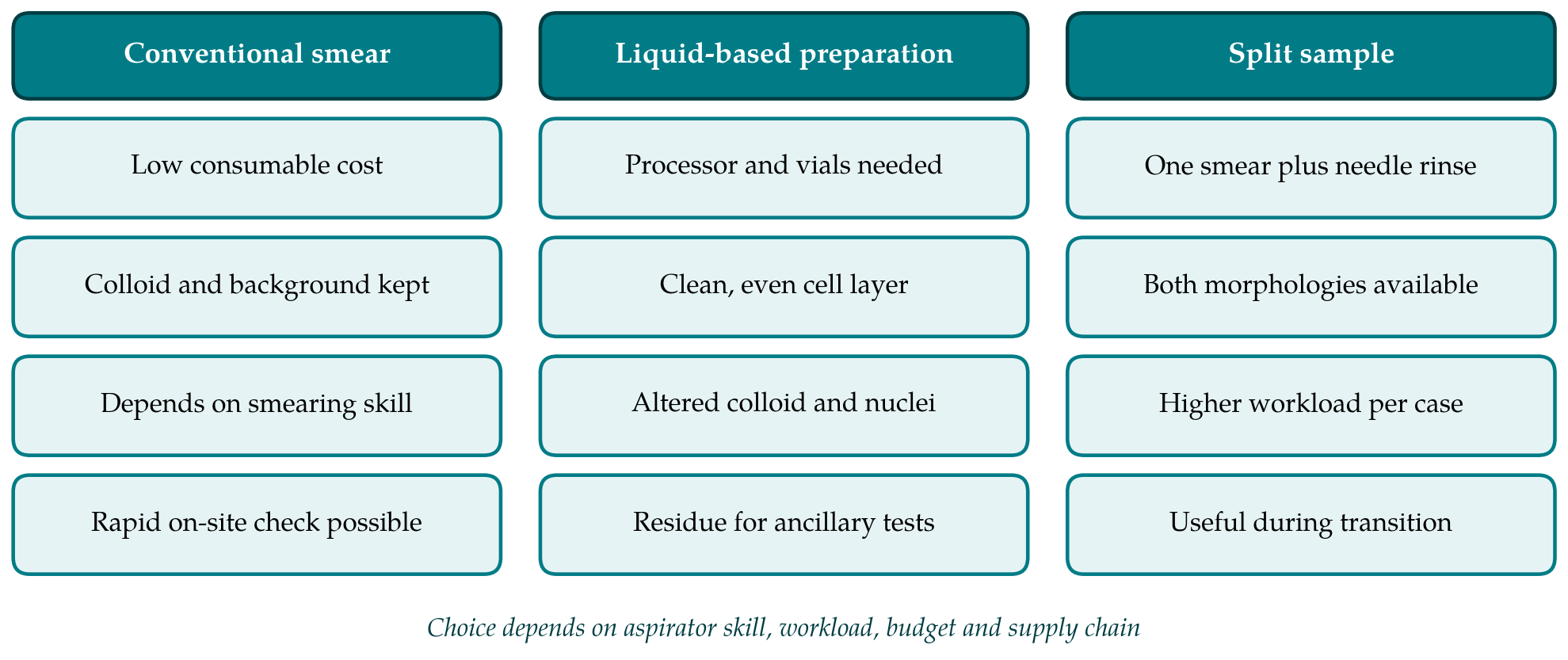
Liquid-based preparation moves slide making into the laboratory. Two commercial systems dominate. One collects cells on a membrane filter from a methanol-based fluid and transfers them to the slide; the other enriches the sample through a density reagent and lets the cells settle from an ethanol-based fluid. Both give a circular deposit of evenly spread, alcohol-fixed cells, read with the Papanicolaou stain, with most of the blood removed (Linder, 1998). Manual substitutes also exist, in which the rinse is fixed in a locally prepared alcohol-based medium, centrifuged and applied to the slide by pipette or cytocentrifuge. Figure 1 sets the main features of the approaches side by side, including the split-sample practice in which a smear is made first and the needle is then rinsed into a vial.
4. How the Morphology Differs
A cytologist trained on smears who first looks at a liquid-based thyroid slide sees a familiar lesion in an unfamiliar dress. The differences were catalogued early for the filter-based method by Michael and Hunter (2000) and for thyroid specifically by Afify et al. (2001) and Cochand-Priollet et al. (2003), and are summarized in Table 1. Colloid, the chief clue to a benign nodule, is reduced in amount and changes form: thin colloid appears as folded sheets likened to tissue paper, and dense colloid as scattered droplets. Large follicular sheets and papillary fronds break into smaller groups, so architecture, on which the distinction between a benign nodule and a follicular-patterned lesion partly rests, is harder to judge.
| Feature | Conventional smear | Liquid-based preparation |
|---|---|---|
| Colloid | Abundant; thin films and thick fragments easily seen | Reduced; folded tissue-paper sheets or dense droplets (Afify et al., 2001) |
| Architecture | Large sheets, intact follicles and papillae | Smaller, tighter groups; fragmented papillae |
| Nuclear detail | Depends on fixation; air-drying artifact common | Uniformly fixed; nucleoli more evident, pseudoinclusions harder to find |
| Background | Blood, lymphocytes and cyst contents retained | Clean; lymphocytes dispersed, blood largely removed |
| Stains | Papanicolaou and Romanowsky | Papanicolaou as routine |
| Screening area | Whole slide, often several slides | One small circular deposit |
| Residual material | None unless a rinse is kept | Vial residue for cell block or molecular tests |
Nuclear features shift as well. Immediate wet fixation gives consistent chromatin and makes nucleoli more conspicuous, which can lend benign follicular cells a worrying look, while the intranuclear pseudoinclusions and pale chromatin of papillary carcinoma may be less obvious than in a good smear (Cochand-Priollet et al., 2003). The background is altered too. Lymphocytes in chronic thyroiditis are scattered and partly lost, and the watery colloid and macrophages of a cyst are thinned out. A clean background helps screening and removes information at once. These are not defects in the sense of errors. They are a different set of appearances that has to be learned, and the nuclear criteria have gained in importance since the reclassification of some encapsulated follicular-patterned tumors as non-invasive neoplasms (Nikiforov et al., 2016).
5. What Comparative Studies Report
The first formal comparison in the thyroid, by Biscotti et al. (1995), was cautious about letting thin-layer slides replace smears, and the retrospective comparison by Afify et al. (2001) likewise favored the conventional preparation, attributing the difference to the artifacts just described. Studies from laboratories with longer experience were more positive. Tulecke and Wang (2004) correlated thin-layer cytology of follicular lesions with histology, Malle et al. (2006) reported satisfactory use of a thin-layer technique, and a multi-year survey of the sedimentation-based system described its routine application to thyroid aspirates (Geers and Bourgain, 2011). Large institutional series have examined how the distribution of diagnostic categories changes with the preparation used (Nagarajan et al., 2015).
The pattern across this literature is that results improve as familiarity grows, which suggests that much of the early deficit lay with the reader and not with the slide. A meta-analysis by Chong et al. (2017) found the two approaches broadly comparable in diagnostic performance, with considerable heterogeneity among the contributing studies and differences between the commercial systems. Heterogeneity is to be expected. The studies differ in whether samples were split or taken separately, in who performed the aspiration, in whether ultrasound guidance was used, and in the reference standard. Split-sample designs in particular favor the smear, which receives the first and richest part of the aspirate.
Adequacy deserves separate comment, because a non-diagnostic result means a repeat procedure and, in a setting where patients travel far, often a patient lost to follow-up. The Bethesda System defines an adequate thyroid aspirate by a minimum number of well-preserved follicular cell groups (Cibas and Ali, 2017), and the same criterion applies to both preparations. Liquid-based processing rescues samples that would have been obscured by blood or ruined by air-drying, yet it cannot create cells that the needle did not collect. Where the non-diagnostic rate is high because of smearing faults, a change of preparation may help; where it is high because of sampling, it will not. A meta-analysis of the Bethesda System shows how widely category rates vary between institutions for reasons unrelated to preparation (Bongiovanni et al., 2012).
6. Costs, Skills and Supply in Resource-Limited Laboratories
The economics are unfavorable to automated liquid-based cytology in a small laboratory. A processor must be bought and maintained, and every case consumes a proprietary vial, filter or reagent and a coated slide. The systems were priced for cervical screening programs with high volumes. A laboratory that handles a modest number of thyroid aspirates spreads the fixed cost over few cases and is exposed to interruptions in supply, import delays and the absence of local service engineers. Analyses of pathology capacity in low-income countries stress that sustainable choices are those that do not depend on a single foreign supplier (Sayed et al., 2018).
Skills pull in the opposite direction. The conventional smear is cheap only if it is made well, and in many hospitals the aspirator is a surgeon, an endocrinologist or a trainee with no instruction in smearing. Ljung (2008) argued that the proper answer to bad smears is to teach people to make good ones, and that liquid-based methods treat the symptom. The argument is persuasive where a cytopathologist performs or attends the aspiration and can assess the material on site. It is less so where samples arrive from distant clinics already spread, dried and broken. For that pattern of referral a vial of fixative that tolerates transport has an obvious practical appeal.
Retraining is a real cost in either direction. A laboratory that converts must expect a period in which readers over-call nucleoli and under-recognize colloid, and the published accounts of pitfalls should be studied before the first case is signed out (Michael and Hunter, 2000). The Bethesda atlas now illustrates liquid-based appearances alongside smears and is the most accessible training resource (Ali and Cibas, 2018). The split-sample approach, with one direct smear and a needle rinse, lets readers compare the two pictures on the same lesion and is a reasonable way to manage the transition, at the price of extra work per case.
7. Ancillary Testing and Manual Alternatives
One advantage of the vial is independent of morphology. Residual fluid can be used for a cell block, for immunocytochemistry or for molecular analysis without a second puncture, and proponents regard this as the strongest argument for liquid-based collection in the thyroid (Rossi et al., 2012; Fadda and Rossi, 2011). Its weight depends on whether such tests are available and affordable locally. Where molecular testing of indeterminate nodules is not offered, the benefit is limited to the occasional immunostain, which a needle rinse collected alongside a conventional smear would serve equally well.
Manual liquid-based methods occupy the middle ground. They need a centrifuge and a locally mixed fixative, both within reach of most laboratories, and they give the clean background and uniform fixation of the commercial systems without the recurring consumable. Their weakness is the lack of standardization: cell loss, uneven deposition and fixative composition vary from one description to the next, and comparative evidence in the thyroid is sparse. They are the option most in need of careful evaluation, because they are the option that resource-limited laboratories can actually sustain.
8. Gaps in the Evidence
Most comparative studies come from well-resourced centers with experienced aspirators, ultrasound guidance and histological follow-up. Their finding of near-equivalence does not transfer automatically to a district hospital where palpation-guided aspirates are smeared by rotating staff. Studies set in such laboratories, with adequacy rate and cost per diagnostic result as outcomes, are few. So are comparisons that include a manual liquid-based arm, or that measure the effect of a short course in smear technique against the purchase of equipment. Until such work is published, recommendations for these settings rest on reasoning from general principles more than on direct evidence.
9. Conclusion
The literature does not show that either preparation is intrinsically more accurate for thyroid aspirates once readers are familiar with it. It does show that the two are not interchangeable without retraining, and that their costs fall in different places: on the skill of the aspirator for the smear, and on equipment and consumables for the liquid-based slide. For a resource-limited laboratory a well-made conventional smear, air-dried and alcohol-fixed, remains a defensible default, and investment in aspiration and smearing technique is likely to be repaid. Liquid-based preparation is most justified where specimens are referred from afar, where smear quality cannot be controlled or where residual material will be used. Manual variants merit systematic study.
Conflict of interest
To be declared by the author(s) before publication.
Funding
To be declared by the author(s) before publication.