1. Introduction
Screening before laser refractive surgery is largely a search for the cornea that will not tolerate it. Unrecognised early keratoconus is the strongest known risk factor for ectasia after surgery (Randleman et al., 2008), and the international consensus on ectatic disease treats posterior elevation and thickness distribution, obtained by tomography, as the core of detection (Gomes et al., 2015). Tomography has a blind spot, however. It describes the front surface of the epithelium and the back surface of the cornea, and says nothing directly about the shape of the stromal surface in between.
The epithelium is not a uniform coat. It thins over stromal elevations and thickens over depressions, so that the front surface is smoother than the stroma beneath. In early keratoconus this compensation can hide a cone from topography. Reinstein et al. (2009) showed that the epithelial thickness profile carries the signature of the cone, a localised zone of thinning surrounded by an annulus of thickening, and later work showed that the pattern can be present in fellow eyes that look normal on conventional analysis (Reinstein et al., 2015). Mapping the epithelium has since moved from specialist ultrasound instruments to widely available optical coherence tomography.
What has not followed is agreement on how the map should be acquired and reported. This article proposes a protocol for doing so in the setting of candidate screening. It is a methodological proposal assembled from the literature on epithelial behaviour and instrument performance. No measurements were made for this article and the protocol has not been validated.
2. Background
The proposal draws on three bodies of knowledge: the normal profile and its variation, the patterns associated with disease and with lens wear, and the properties of the instruments.
2.1. The normal profile
Very high-frequency digital ultrasound first provided maps over a wide diameter. In normal eyes the central epithelium averages a little over 50 micrometres, and it is thinner superiorly than inferiorly, an asymmetry attributed to the action of the upper lid (Reinstein et al., 2008). Optical coherence tomography gave the same qualitative picture and added reference data by sex and age (Kanellopoulos and Asimellis, 2013; Yang et al., 2014). The implication for a protocol is that a normal map is not flat, and that an index of asymmetry must be judged against a normal superior to inferior gradient and not against zero.
2.2. Patterns that matter in screening
In keratoconus the thinnest epithelium lies over the cone, usually inferotemporal to the centre, with thickening around it; total and stromal thickness maps show the cone more starkly once the epithelium is removed from the calculation (Reinstein et al., 2010). Li et al. (2012) derived pattern-based indices from optical coherence tomography that separated keratoconic from normal eyes, Silverman et al. (2014) built a classifier on epithelial variables, and Temstet et al. (2015) examined the form fruste stage. Contact lens warpage gives the opposite relation between curvature and epithelium: where topography steepens, the epithelium is thick and not thin (Schallhorn et al., 2017). Dry eye has been associated with altered epithelial thickness as well (Kanellopoulos and Asimellis, 2014). A map is therefore read as a pattern in relation to curvature, and a single thickness value has little meaning.
2.3. Instruments
Optical coherence tomography (Huang et al., 1991) measures the epithelium by segmenting the air to tear interface and the boundary at Bowman's layer. The precorneal tear film is included in the optical measurement, whereas immersion ultrasound excludes it. Instruments differ in wavelength, axial resolution, scan pattern, mapped diameter and segmentation software, and their readings are not interchangeable. Repeatability in normal and post-LASIK eyes has been reported as good for a Fourier-domain device (Ma et al., 2013), but precision is poorer toward the periphery and in irregular corneas, which are the eyes of most interest.
3. Sources of Variability
Table 1 lists the influences that a protocol has to control and the measure proposed for each. They fall into three groups. Some change the epithelium itself, such as recent contact lens wear and ocular surface disease. Some change what the instrument sees without changing the tissue, such as an unstable tear film, residue from viscous drops or a decentred scan. The last group lies in the device and its software, including segmentation errors and differences between instruments.
| Source | Effect on the map | Proposed control |
|---|---|---|
| Contact lens wear | Regional thickening or thinning that mimics or masks disease | Record lens type and last wear; repeat after a lens-free interval until stable |
| Drops and contact tests | Surface disturbance from anaesthetic, dye, tonometry or gonioscopy | Map before any drop or contact procedure at the visit |
| Tear film | Optical thickness includes the tear layer; break-up adds noise | Scan shortly after a natural blink; note surface disease |
| Centration and fixation | Shifts sector values and the apparent site of thinning | Fix the reference point and reject decentred scans |
| Eyelids and lashes | Missing superior data; pressure distorts the cornea | Lift the lid against the orbital rim without pressing on the globe |
| Segmentation | Boundary errors produce false focal thin or thick spots | Inspect the B-scans behind every abnormal sector |
| Device and software | Systematic offsets between instruments and versions | One device per patient pathway; record model and software version |
4. The Proposed Protocol
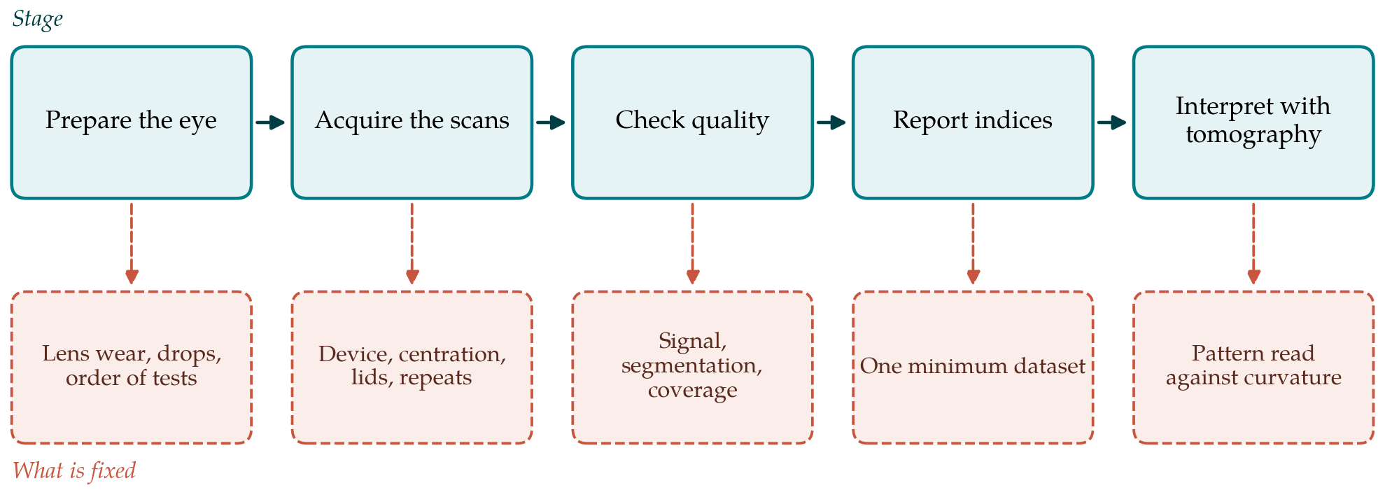
The protocol has five stages, set out in Figure 1 with the element that each stage fixes. It is written to be independent of manufacturer. Where a clinic's instrument cannot provide an element, the clinic records that fact instead of substituting a different quantity under the same name.
4.1. Stage 1: Preparing the eye
Mapping is placed first in the order of tests, before tonometry, anaesthetic, fluorescein, dilation or any procedure that touches the cornea. Contact lens history is recorded by lens type, wearing pattern and time since last wear. The literature does not support one lens-free interval for all wearers, and the protocol does not invent one. It asks instead for stability: lens wearers are mapped, mapped again after a further lens-free period chosen by the clinic's existing policy for refraction and topography, and accepted when the two maps agree within the clinic's own repeatability limit. Signs of ocular surface disease are noted, and marked surface disease is treated before the map is used for screening.
4.2. Stage 2: Acquisition
The same instrument, scan pattern and mapped diameter are used for every candidate, and the widest map the device offers is preferred because the annulus of thickening around a cone may lie outside a small central zone. The reference point for centration, whether pupil centre or corneal vertex, is fixed in the protocol and stated in reports. The patient blinks naturally and the scan is taken within a few seconds. If the upper lid obscures the superior cornea it is lifted against the orbital rim. Three acceptable scans are acquired per eye, with the patient sitting back between them, so that within-session repeatability can be calculated for the individual and not assumed from published averages.
4.3. Stage 3: Quality control
A scan is accepted only if the manufacturer's signal quality index is above the recommended level, the map is complete in all sectors to be reported, centration is within the instrument's tolerance and the segmentation lines follow the anatomical boundaries on inspection of the cross-sections. The last check matters most. Automated segmentation can fail over scars, in the periphery and where the tear film is irregular, and a focal abnormality on the map that does not correspond to a visible change in the B-scan is treated as artefact. Rejected scans are counted and the count is recorded.
4.4. Stage 4: A minimum reported dataset
Published studies report different indices, which makes thresholds hard to compare. The protocol specifies a short common set to be reported from the mean of the accepted scans: central thickness; minimum and maximum thickness with their locations; the difference between minimum and maximum; the superior to inferior difference; the standard deviation across the map; and the mapped diameter. The within-session standard deviation of each index is reported beside it. Indices that depend on proprietary software may be added under their own names. The colour scale and step are fixed so that maps from different visits can be compared by eye.
4.5. Stage 5: Interpretation with tomography
The map is never read alone. It is placed beside the anterior curvature, posterior elevation and pachymetry maps, and the reader answers one question first: whether the thinnest epithelium coincides with the steepest curvature and the thinnest stroma. Coincident thinning with a surrounding thick zone supports ectasia. Thick epithelium over an area of steepening supports warpage or another non-ectatic cause. A map that is normal in pattern does not clear an eye with abnormal tomography, and an abnormal map in an eye with normal tomography is a reason for further assessment, not a diagnosis.
A repeat or defer rule completes the protocol. If the three scans disagree by more than the clinic's repeatability limit, if the pattern is plausibly due to lens wear or surface disease, or if map and tomography conflict, the examination is repeated at a later visit under the same conditions before a surgical decision is taken.
5. Setting Local Limits
The protocol refers several times to a clinic's own repeatability limit, and it does so on purpose. Published cut-offs come from particular instruments, software versions and populations, and carrying them to another setting assumes an equivalence that has not been shown. A clinic adopting the protocol would first establish its own precision, by repeated measurement of a set of normal eyes by the operators who will perform screening, analysed with within-subject standard deviation and limits of agreement in the manner described by Bland and Altman (1986) and, for ophthalmic instruments specifically, by McAlinden et al. (2011). Operator and session effects should be included, since real screening involves both.
6. Limitations and What Remains to Be Shown
This article proposes a protocol and reports no test of it. Whether the controls in Table 1 reduce variability in practice, and by how much, is an empirical matter. So is the larger question of whether a standardised map, added to tomography, improves the detection of eyes at risk. Answering it requires a diagnostic accuracy study with a defined reference standard, consecutive enrolment and reporting to the STARD guideline (Bossuyt et al., 2015). The reference standard is itself a difficulty, because early ectasia has no independent gold standard and progression over time is the nearest substitute.
Other limits should be stated. The protocol does not resolve differences between devices and makes no claim that indices transfer from one to another. It does not set a lens-free interval. It addresses screening of untreated eyes; mapping after surgery, where the epithelium remodels in response to the ablation, raises separate questions. Finally, a protocol adds chair time, and a clinic would need to judge whether three scans per eye and inspection of cross-sections are sustainable in its setting.
7. Conclusion
Epithelial thickness mapping can show what topography hides, but only if the map reflects the tissue and not the conditions of the examination. The five-stage protocol proposed here fixes the order of tests, the acquisition settings, the quality checks, a minimum set of reported indices and the way the map is read against tomography, and it asks each clinic to derive its own repeatability limits before applying thresholds. It is offered for trial and criticism. Its usefulness will be decided by repeatability and diagnostic accuracy studies that have yet to be carried out.
Conflict of interest
To be declared by the author(s) before publication.
Funding
To be declared by the author(s) before publication.