1. Introduction
Lumpy skin disease (LSD) is caused by lumpy skin disease virus (LSDV), a large double-stranded DNA virus placed with sheeppox virus and goatpox virus in the genus Capripoxvirus. The three viruses are closely related, cannot be told apart by routine serology, and cross-protect to a degree that has shaped vaccination policy for decades (Babiuk et al., 2008). Affected cattle develop fever, enlarged superficial lymph nodes and firm, circumscribed skin nodules that may cover the whole body. Milk yield falls, bulls may become temporarily or permanently infertile, pregnant cows may abort, and hides are permanently damaged. Many infections are mild or inapparent, and deaths are usually few, yet the cumulative loss of production and the trade restrictions that follow an outbreak make the disease one of the costlier infections of cattle (Tuppurainen and Oura, 2012).
The disease was first recognised in Zambia in 1929 and for decades was regarded as an African problem. It reached Israel in 1989, spread through the Middle East, and entered Turkey in 2013 and south-east Europe and Russia from 2015. In 2019 it was reported from Bangladesh, India and China, and within three years it had been recorded in most countries of South and South-East Asia (Roche et al., 2020). The cattle and buffalo populations now exposed are among the largest in the world and are kept mostly by smallholders. This review considers what is known about how the virus spreads, how well the available vaccines work, and what these two bodies of knowledge imply for control in Asia.
2. Scope and Approach
This is a narrative review of the published literature. It draws on experimental transmission studies, vaccine trials and field evaluations, outbreak reports from Asian countries, genomic analyses of field strains, and the guidance documents issued by the Food and Agriculture Organization of the United Nations and the European Food Safety Authority. Sources were chosen for their relevance to the three questions set out above and not through a formal systematic search. Older African and Middle Eastern work is included wherever it remains the main evidence on a point, as it does for most of the vector studies.
3. The Virus and the Course of Infection
The LSDV genome is about 151 kilobase pairs long and encodes genes for host range and immune evasion that are disrupted in the sheep and goat viruses (Tulman et al., 2001). The classical account of the disease, drawn from southern African outbreaks, remains accurate: an incubation period of one to four weeks, fever, nodules that involve the full thickness of the skin and later form necrotic plugs, and lesions in the mouth, respiratory tract and udder (Tuppurainen and Oura, 2012). After experimental inoculation only a proportion of animals develop generalised disease. Virus is abundant in skin nodules and persists there for weeks, and it is found at lower levels and for shorter periods in blood and in nasal, oral and ocular secretions (Tuppurainen et al., 2017a). Bulls may shed virus in semen for a prolonged period after recovery (Irons et al., 2005). The skin is therefore the main source of virus for whatever carries it to the next animal.
Laboratory confirmation rests on detection of capripoxvirus DNA by real-time polymerase chain reaction. Skin nodules and scabs are the samples of choice. Antibody tests are less sensitive, particularly in mildly affected and vaccinated animals, because protection against poxviruses depends largely on cell-mediated immunity (Tuppurainen et al., 2017a).
Clinical recognition is straightforward in a severe case and difficult in a mild one. A few small nodules on the neck, perineum or udder are easily overlooked or attributed to insect bites, pseudo-lumpy skin disease caused by bovine herpesvirus 2, dermatophilosis or demodicosis. The field manual prepared for veterinarians recommends that any cattle with multiple firm skin nodules and fever in an area at risk be sampled (Tuppurainen et al., 2017b). Mildly affected animals matter for control because they are the ones most likely to be moved or sold while infectious.
4. Emergence and Spread in Asia
Figure 1 summarises the widening range of the disease. The first Asian outbreaks outside the Middle East were recognised in 2019. In Bangladesh the disease appeared in the south-east of the country in the middle of that year (Badhy et al., 2021). In India the first confirmed outbreaks were in the eastern state of Odisha in August 2019 (Sudhakar et al., 2020). China reported its first outbreak in the same month in Xinjiang, close to the border with Kazakhstan (Lu et al., 2021).
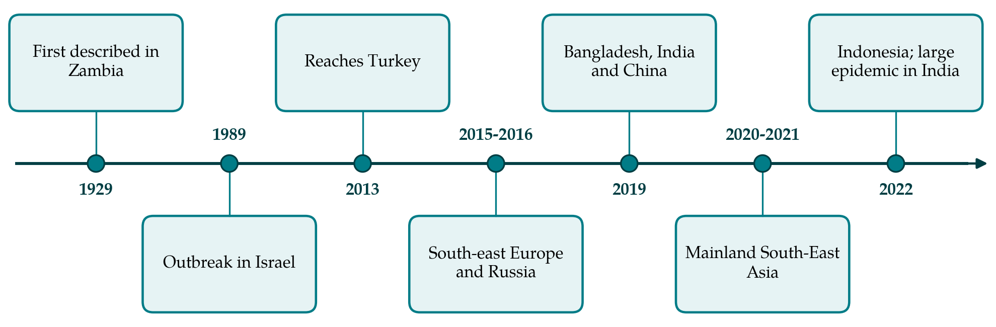
From these footholds the disease moved quickly. Vietnam reported outbreaks in its northern provinces in late 2020 (Tran et al., 2021), Thailand followed in 2021, and Hong Kong recorded clinical disease in feral cattle (Flannery et al., 2022). A risk assessment prepared for the region had anticipated this course, identifying informal cross-border trade in live cattle and the density of susceptible animals as the main drivers (Roche et al., 2020). India experienced a far larger epidemic in 2022, concentrated in the western and northern states.
Genomic studies show that Asia received the virus by more than one route. The strains found in Bangladesh and India in 2019 resembled long-established field strains of African origin and were distinct from those then circulating in Europe and the Middle East (Badhy et al., 2021; Sudhakar et al., 2020). The strains detected in China, Vietnam, Thailand and Hong Kong belong to a different group: they are recombinants that combine sequence from field virus with sequence characteristic of live vaccine strains (Flannery et al., 2022). Viruses of this kind were first described in Russia, in regions where the homologous vaccine was not in official use (Sprygin et al., 2018; Kononov et al., 2019).
5. Transmission Dynamics
The seasonal pattern of outbreaks, with peaks in warm and wet months and a decline in cold or dry periods, pointed to arthropod transmission long before it was shown experimentally. Table 1 lists the routes that have been examined and the strength of the evidence for each.
| Route | State of the evidence | Representative sources |
|---|---|---|
| Stable flies (Stomoxys calcitrans) | Mechanical transmission to cattle shown experimentally | Sohier et al., 2019 |
| Horseflies (Haematopota spp.) | Mechanical transmission shown experimentally | Sohier et al., 2019 |
| Mosquitoes (Aedes aegypti) | Mechanical transmission shown for several days after feeding | Chihota et al., 2001 |
| Other biting insects | Attempts with several species did not transmit; virus detected in some | Chihota et al., 2003 |
| Hard ticks | Mechanical transmission by male Rhipicephalus appendiculatus shown | Tuppurainen et al., 2013 |
| Direct contact | Inefficient when vectors are excluded | Carn and Kitching, 1995 |
| Semen | Prolonged shedding by bulls documented | Irons et al., 2005 |
5.1. Vectors
No arthropod has been shown to support replication of LSDV. Transmission is mechanical: an insect interrupted while feeding on a nodule carries virus on its mouthparts to the next host. The first experimental demonstration used the mosquito Aedes aegypti, which remained able to infect cattle for several days after feeding on lesions (Chihota et al., 2001). The same group could not show transmission with the stable fly or with several other biting insects under their conditions (Chihota et al., 2003), a negative result that stood for years against strong field suspicion of Stomoxys. Later experiments with larger numbers of flies and donors with abundant lesions did achieve transmission by stable flies and by horseflies (Sohier et al., 2019). Male ixodid ticks, which move between hosts in search of females, can also carry the virus (Tuppurainen et al., 2013).
These studies establish which vectors are capable of transmission. They do not show which are responsible for it in a given place, and almost none of the work was done with Asian vector species or in Asian husbandry systems (Sprygin et al., 2019). Stable flies breed in manure and decaying fodder around housed cattle. Tabanids and mosquitoes depend on standing water and are strongly seasonal in monsoon climates. The relative weight of each is likely to differ between a tethered smallholder herd, a peri-urban dairy colony and free-grazing cattle, and this remains an open field question.
5.2. Contact, fomites and animal movement
When infected and susceptible cattle were housed together in insect-proof conditions, transmission was rare, which led to the view that contact alone does not sustain an outbreak (Carn and Kitching, 1995). Shared water troughs, contaminated feed and reuse of needles during mass treatment or vaccination are plausible routes that have been little studied. Flying insects account for spread over short distances between neighbouring herds. Jumps over long distances are attributed to the transport of infected animals, including those incubating the disease or carrying mild lesions hidden under the hair coat (EFSA, 2015). In much of Asia cattle move along trade routes that cross several borders without veterinary inspection, which fits the observed pattern of sudden appearance far from known foci.
6. Vaccination
Vaccination is the only measure that has reliably stopped LSD epidemics. All vaccines in widespread use are live attenuated capripoxviruses, and they fall into two groups.
6.1. Homologous and heterologous vaccines
Homologous vaccines contain attenuated LSDV, most often derived from the South African Neethling strain. Heterologous vaccines contain attenuated sheeppox or goatpox virus and were adopted in the Middle East and parts of Asia where the sheep and goat diseases were already present and the use of live LSDV was not permitted. Direct comparison favours the homologous product. In a randomised field study in Israeli dairy herds, a Neethling vaccine protected markedly better than a sheeppox vaccine given at ten times the sheep dose (Ben-Gera et al., 2015). In an Ethiopian challenge experiment, a goatpox vaccine protected cattle while a Neethling vaccine and a strain marketed as a sheep and goat pox vaccine did not, a result that drew attention to differences between manufacturers and seed strains as much as between virus species (Gari et al., 2015). The field manual issued by the Food and Agriculture Organization accordingly advises homologous vaccine where it is licensed and available (Tuppurainen et al., 2017b).
The strongest field evidence comes from the Balkans, where coordinated mass vaccination with Neethling vaccines across several countries was followed by the disappearance of outbreaks, and analysis of the epidemic data indicated high effectiveness (Klement et al., 2020). A minority of cattle given Neethling vaccines develop a local reaction at the injection site, a short fever and drop in milk, and occasionally small generalised nodules, the so-called Neethling response. Owners who meet these reactions without warning may refuse later rounds.
6.2. Recombinant strains and vaccine quality
The discovery of field viruses carrying vaccine-like sequence changed the discussion of vaccine safety. Recombination between poxviruses requires two viruses to infect the same cell, and the Russian findings were first read as evidence that a vaccine strain had recombined with field virus in vaccinated cattle (Sprygin et al., 2018). The origin of these strains is still debated in the literature, and contamination or mixing of strains during vaccine manufacture has been proposed as an alternative. Whatever their origin, recombinant viruses now predominate in East and South-East Asia, and they complicate diagnosis, because assays designed to separate vaccine from field virus by a single genetic marker can misclassify them (Kononov et al., 2019). The practical lessons are that vaccines should come from manufacturers whose seed virus and batches are independently tested, and that no licensed vaccine yet allows infected animals to be distinguished from vaccinated ones by serology (Tuppurainen et al., 2017a).
7. Control in Asian Settings
European control combined early detection, movement restrictions, stamping out in some countries and region-wide vaccination, supported by compensation and reliable animal identification (EFSA, 2015). Few of these conditions hold where the disease is now spreading. Culling of cattle is unacceptable or unlawful in parts of South Asia, herds are small and numerous, animals graze communally, and veterinary services are stretched. Control therefore depends almost entirely on vaccination coverage, and coverage depends on vaccine supply, cold chain, the cost to the owner and trust in the product. Countries that began with goatpox or sheeppox vaccines because these were produced domestically face the question of whether, and when, to change to homologous vaccines.
Supporting measures have a modest but real place. Movement of cattle from affected areas, and especially sale of visibly sick animals, should be discouraged, although enforcement at informal markets is difficult. Insect control around animals may reduce biting pressure, but there is little evidence that insecticides or repellents alter the course of an outbreak, and they should not be presented to farmers as a substitute for vaccination (Tuppurainen et al., 2017b). Affected animals need supportive care and treatment of secondary infection, and clean needles should be used for every animal. Since the disease does not infect people, public communication can concentrate on animal health and on the safety of milk and meat.
Surveillance is the weakest part of control in much of the region. Outbreaks are reported by owners or field staff on clinical grounds, laboratory confirmation is available at a few national centres, and sequence data come from a small number of samples. A regional picture of which strains circulate where, and of how they respond to the vaccines in use, requires routine sharing of samples and genomic information between neighbouring countries, which the risk assessment for the region identified as a priority (Roche et al., 2020).
8. Gaps and Future Work
Several questions are specific to Asia. The vector species responsible for transmission in monsoon and tropical climates have not been identified by field studies, and overwintering of the virus between seasons is unexplained. Water buffalo are susceptible but seem to be affected less often and less severely than cattle; their part in maintaining infection where they are kept alongside cattle is unknown. The same is true of wild bovids in the region. On the vaccine side, the comparative effectiveness of the goatpox, sheeppox and Neethling products actually used in Asian programmes has not been measured against the recombinant strains now circulating. Inactivated vaccines and vaccines with markers that permit differentiation of infected from vaccinated animals are under development and would remove two of the main obstacles to regional control (Tuppurainen et al., 2017a).
9. Conclusion
Lumpy skin disease has become endemic over a large part of Asia within a few years, carried by cattle movements over distance and by biting arthropods locally. The experimental literature identifies stable flies, horseflies, mosquitoes and ticks as competent mechanical vectors, but leaves their field importance in Asian systems unresolved. Homologous live vaccines of assured quality, applied at high coverage, have the best record, and the recombinant strains now circulating make vaccine quality control and careful diagnostic interpretation more necessary than before. Regional coordination of vaccination and of information on circulating strains offers the most realistic route to reducing the burden of the disease.
Conflict of interest
To be declared by the author(s) before publication.
Funding
To be declared by the author(s) before publication.